We are very proud of having solved most of the structures of septins deposited in the PDB!
These include single isolated G-domains for many different septins (including those from humans, Schistosoma and Chlamydomonas) as well dimeric and trimeric complexes and isolated coiled-coil domains. More recently, we have determined the first cryo-EM septin structure, the SEPT2-6-7 trimeric complex.
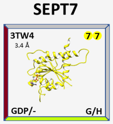
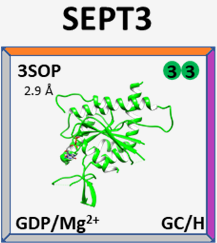
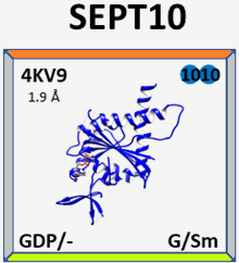
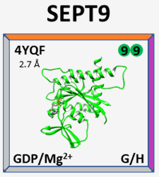
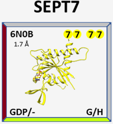
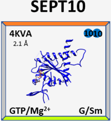
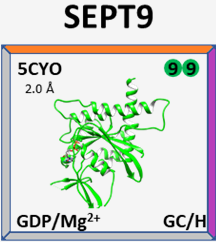
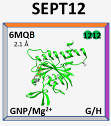
Below, there is a gallery of our released structures. As an example, we show the highest resolution septin structure solved to date (SEPT7 at 1.7 Å resolution):
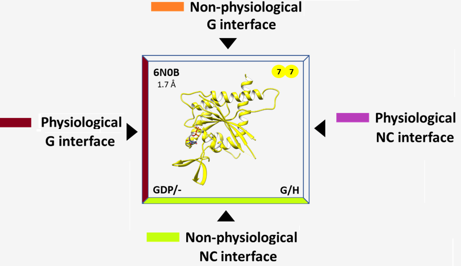
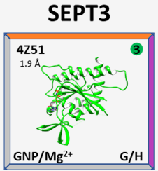
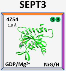
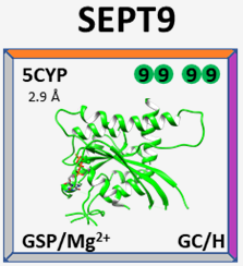
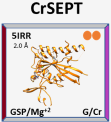
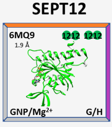
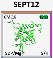
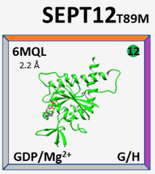
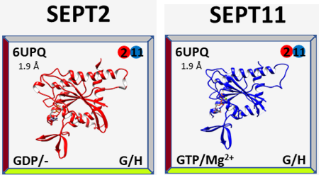
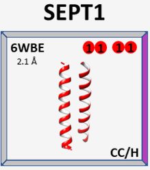
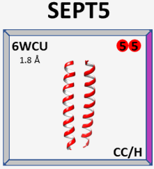
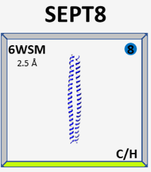
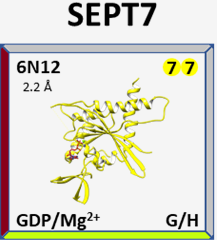
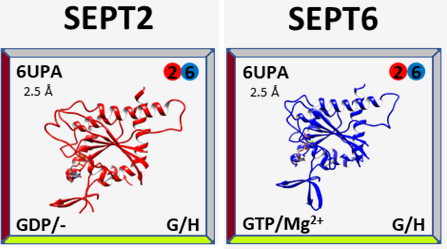
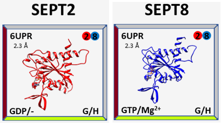
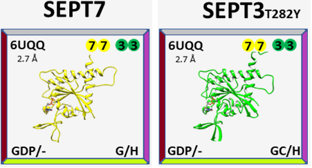
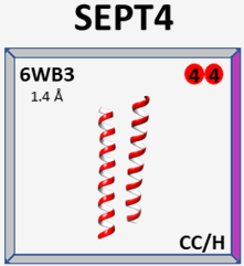
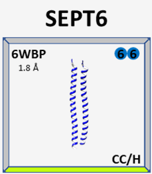
Each subunit of a structure is represented in a square and is colored according to its septin group. Clicking on them will redirect you to their PDB pages. PDB entry and resolution, asymmetric unit composition, nucleotide/magnesium binding and crystallized domain(s)/taxonomy are displayed. Corners are colored according to the interfaces found in the structure, classified in physiological (i.e., based on the canonical models) or not.
8DKT
8SGD
8FWP
8SJJ
9BHT
9BHW